
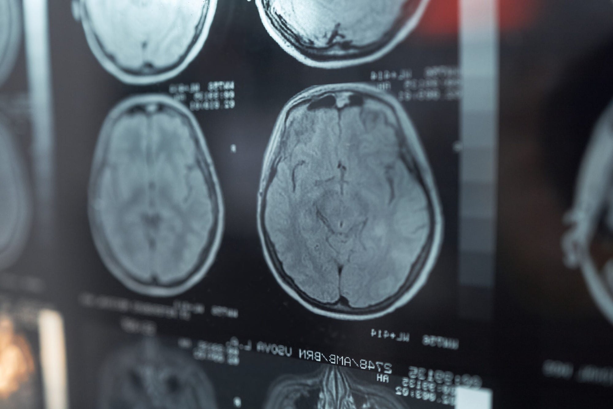
A damaged patch of tissue in the adult brain can trigger a repair program in neighbouring supporting cells, one that was previously thought impossible. The cells stretch out, divide, and then move their newly formed nuclei into the emptied area. The process was observed in living mice by researchers at the University of Zurich.
At the centre of the study were astrocytes, star-shaped glial cells that support neurons and maintain a healthy environment in brain tissue. The scientists selectively destroyed small groups of these cells in the somatosensory cortex of mice. To do this, they used antibodies against aquaporin-4, a water channel found mainly on astrocytes. Similar antibodies appear in people with neuromyelitis optica, an autoimmune disease in which the immune system attacks precisely these cells.
For a long time it was believed that the adult brain cannot make up for a substantial loss of astrocytes. However, observations made with two-photon microscopy over periods of up to 60 days showed the opposite. Surviving astrocytes did not form a scar around the damage; instead, they entered a temporary regenerative state. Their processes lengthened and extended towards the centre of the affected area, reaching maximum length at around the fifth day. By that point, the territory occupied by a single cell had grown by 70 percent on average.
The cells then began to divide. About 60% of the astrocytes surrounding the damage had divided within two months. Some went through several rounds of division, producing up to four daughter cells. The most unusual part of the process came next: instead of dividing right away, the parent cell temporarily contained several nuclei. These new nuclei then travelled along long processes into the empty areas. In the words of Bruno Weber, who led the study, the cells "send the newly formed nuclei of their daughter cells sliding over large distances to repopulate the damaged part of the brain and re-knit the network of astrocytes."
Most of the nuclei moved within the first two weeks. Some covered a distance of more than 100 micrometres. They moved unevenly, with pauses, reaching speeds of up to 5.6 micrometres per hour. These physical changes were accompanied by a temporary reorganisation of gene activity: genes associated with cellular plasticity and structural remodelling were switched on. The peak of this repair program came on the fifth day, and by the seventeenth day its traces were no longer detectable.
The astrocytes' response depended on the nature and scale of the damage. Smaller lesions, similar to a stroke, prompted division, but recovery remained incomplete. The strongest response, involving division and the movement of nuclei over long distances, was triggered by extensive loss of cells.
The researchers also examined brain samples from people with confirmed neuromyelitis optica. In the affected areas they found astrocytes with similar features: elongated, oriented towards the damage, sometimes with several nuclei and signs of division. The authors of the work, published in the journal Nature Neuroscience, stress that this does not prove the same process occurs in humans, since the human disease involves chronic inflammation and broader tissue damage, which was not present in the mouse model.


Letters